Page Not Found
Page not found. Your pixels are in another canvas.
A list of all the posts and pages found on the site. For you robots out there is an XML version available for digesting as well.
Page not found. Your pixels are in another canvas.
About me
This is a page not in th emain menu
Published:
This post will show up by default. To disable scheduling of future posts, edit config.yml and set future: false.
Published:
This is a sample blog post. Lorem ipsum I can’t remember the rest of lorem ipsum and don’t have an internet connection right now. Testing testing testing this blog post. Blog posts are cool.
Published:
This is a sample blog post. Lorem ipsum I can’t remember the rest of lorem ipsum and don’t have an internet connection right now. Testing testing testing this blog post. Blog posts are cool.
Published:
This is a sample blog post. Lorem ipsum I can’t remember the rest of lorem ipsum and don’t have an internet connection right now. Testing testing testing this blog post. Blog posts are cool.
Published:
This is a sample blog post. Lorem ipsum I can’t remember the rest of lorem ipsum and don’t have an internet connection right now. Testing testing testing this blog post. Blog posts are cool.
Published in Advanced Materials Technologies, 2019
Laser writing of materials is normally performed by the sequential scanning of a single focused beam across a sample. This process is time-consuming and it can severely limit the throughput of laser systems in key applications such as surgery, microelectronics, or manufacturing. A parallelization strategy based on ultrasound waves in a liquid to diffract light into multiple beamlets is reported. Adjusting amplitude, frequency, or phase of ultrasound allows tunable multifocus distributions with sub-microsecond control. When combined with sample translation, the dynamic splitting of light leads to high-throughput laser processing, as demonstrated by locally modifying the morphological and wettability properties of metals, polymers, and ceramics. The results illustrate how acousto-optofluidic systems are universal tools for fast multifocus generation, with potential impact in fields such as imaging or optical trapping.
Recommended citation: Alessandro Zunino, Salvatore Surdo, and Martí Duocastella. “Dynamic Multifocus Laser Writing with Acousto-Optofluidics”. Advanced Materials Technologies 4.12 (2019), pp. 1–7 https://doi.org/10.1002/admt.201900623
Published in Biophysical Chemistry, 2019
Alpha-Synuclein (AS) is the protein playing the major role in Parkinson’s disease (PD), a neurological disorder characterized by the degeneration of dopaminergic neurons and the accumulation of AS into amyloid plaques. The aggregation of AS into intermediate aggregates, called oligomers, and their pathological relation with biological membranes are considered key steps in the development and progression of the disease. Here we propose a multi-technique approach to study the effects of AS in its monomeric and oligomeric forms on artificial lipid membranes containing GM1 ganglioside. GM1 is a component of functional membrane micro-domains, called lipid rafts, and has been demonstrated to bind AS in neurons. With the aim to understand the relation between gangliosides and AS, here we exploit the complementarity of microscopy (Atomic Force Microscopy) and neutron scattering (Small Angle Neutron Scattering and Neutron Reflectometry) techniques to analyze the structural changes of two different membranes (Phosphatidylcholine and Phosphatidylcholine/GM1) upon binding with AS. We observe the monomer- and oligomer-interactions are both limited to the external membrane leaflet and that the presence of ganglioside leads to a stronger interaction of the membranes and AS in its monomeric and oligomeric forms with a stronger aggressiveness in the latter. These results support the hypothesis of the critical role of lipid rafts not only in the biofunctioning of the protein, but even in the development and the progression of the Parkinson’s disease.
Recommended citation: Fabio Perissinotto, Valeria Rondelli, Pietro Parisse, Nicolò Tormena, Alessandro Zunino, László Almásy, Dániel Géza Merkel, László Bottyán, Szilárd Sajti, and Loredana Casalis. “GM1 Ganglioside role in the interaction of Alpha-synuclein with lipid membranes: Morphology and structure”. Biophysical Chemistry 255 (2019), p. 106272 https://doi.org/10.1016/j.bpc.2019.106272
Published in Journal of Physics: Photonics, 2020
Acoustic waves in an optical medium cause rapid periodic changes in the refraction index, leading to diffraction effects. Such acoustically controlled diffraction can be used to modulate, deflect, and focus light at microsecond timescales, paving the way for advanced optical microscopy designs that feature unprecedented spatiotemporal resolution. In this article, we review the operational principles, optical properties, and recent applications of acousto-optic (AO) systems for advanced microscopy, including random-access scanning, ultrafast confocal and multiphoton imaging, and fast inertia-free light-sheet microscopy. As AO technology is reaching maturity, designing new microscope architectures that utilize AO elements is more attractive than ever, providing new exciting opportunities in fields as impactful as optical metrology, neuroscience, embryogenesis, and high-content screening.
Recommended citation: Martí Duocastella et al 2021 J. Phys. Photonics 3 012004 https://doi.org/10.1088/2515-7647/abc23c
Published in Acta Imeko, 2020
The high versatility of laser direct-write (LDW) systems offers remarkable opportunities for Industry 4.0. However, the inherent serial nature of LDW systems can seriously constrain manufacturing throughput and, consequently, the industrial scalability of this technology. Here we present a method to parallelise LDWs by using acoustically shaped laser light. We use an acousto-optofluidic (AOF) cavity to generate acoustic waves in a liquid, causing periodic modulations of its refractive index. Such an acoustically controlled optical medium diffracts the incident laser beam into multiple beamlets that, operating in parallel, result in enhanced processing throughput. In addition, the beamlets can interfere mutually, generating an intensity pattern suitable for processing an entire area with a single irradiation. By controlling the amplitude, frequency, and phase of the acoustic waves, customised patterns can be directly engraved into different materials (silicon, chromium, and epoxy) of industrial interest. The integration of the AOF technology into an LDW system, connected to a wired-network, results into a cyber-physical system (CPS) for advanced and high-throughput laser manufacturing. A proof of concept for the computational ability of the CPS is given by monitoring the fidelity between a physical laser-ablated pattern and its digital avatar. As our results demonstrate, the AOF technology can broaden the usage of lasers as machine tools for industry 4.0
Recommended citation: Salvatore Surdo, Alessandro Zunino, Alberto Diaspro, and Martí Duocastella. “Acoustically-shaped laser: a machining tool for Industry 4.0”. ACTA IMEKO 9.4 (2020), p. 60. https://doi.org/10.21014/acta_imeko.v9i4.740
Published in Frontiers in Physics, 2021
Acoustic waves in an optical medium cause rapid periodic changes in the refraction index, leading to diffraction effects. Such acoustically controlled diffraction can be used to modulate, deflect, and focus light at microsecond timescales, paving the way for advanced optical microscopy designs that feature unprecedented spatiotemporal resolution. In this article, we review the operational principles, optical properties, and recent applications of acousto-optic (AO) systems for advanced microscopy, including random-access scanning, ultrafast confocal and multiphoton imaging, and fast inertia-free light-sheet microscopy. As AO technology is reaching maturity, designing new microscope architectures that utilize AO elements is more attractive than ever, providing new exciting opportunities in fields as impactful as optical metrology, neuroscience, embryogenesis, and high-content screening.
Recommended citation: Callegari, F., Le Gratiet, A., Zunino, A., Mohebi, A., Bianchini, P., & Diaspro, A. (2021). Polarization Label-Free Microscopy Imaging of Biological Samples by Exploiting the Zeeman Laser Emission. Frontiers in Physics, 9, 758880. https://doi.org/10.3389/fphy.2021.758880
Published in ACS Photonics, 2021
Light-sheet microscopes have become the tool of choice for volumetric imaging of large samples. Based on a wide-field acquisition scheme, they are capable of optical sectioning at diffraction-limited resolution and minimal overall photodamage. Unfortunately, traditional architectures are limited in speed because 3D images are collected by either sample translation or synchronized movement of both light-sheet and detection objective lens. A promising solution avoiding slow mechanical movements is to extend the depth-of-field of the microscope and moving only the light-sheet. However, this normally comes at the cost of losing light and contrast, compromising the signal-to-noise ratio of the images. Here, we propose an innovative technique devoted to restoring the quality of the images, while preserving the speed of extended depth-of-field microscopes. It is based on generating a stack of parallel light-sheets using a pair of orthogonal acousto-optic deflectors, enabling the simultaneous illumination of different sample planes. Given the extended depth-of-field, all such planes appear in focus and can be acquired in a superimposed single frame. By applying a single-step inversion algorithm, we can decode a stack of frames into a volumetric image whose signal-to-noise ratio and contrast are greatly enhanced. We provide a detailed theoretical framework of the method and demonstrate its feasibility with volumetric images of kidney cell spheroids.
Recommended citation: Alessandro Zunino, Francesco Garzella, Alberta Trianni, Peter Saggau, Paolo Bianchini, Alberto Diaspro, and Martí Duocastella. “Multiplane Encoded Light-Sheet Microscopy for Enhanced 3D Imaging”. ACS Photonics 8.11 (2021), pp. 3385–3393 https://doi.org/10.1021/acsphotonics.1c01401
Published in Journal of Colloid and Interface Science, 2022
Hypothesis Polymeric anisotropic soft microparticles show interesting behavior in biological environments and hold promise for drug delivery and biomedical applications. However, self-assembly and substrate-based lithographic techniques are limited by low resolution, batch operation or specific particle geometry and deformability. Two-photon polymerization in microfluidic channels may offer the required resolution to continuously fabricate anisotropic micro-hydrogels in sub-10 µm size-range. Experiments Here, a pulsed laser source is used to perform two-photon polymerization under microfluidic flow of a poly(ethylene glycol) diacrylate (PEGDA) solution with the objective of realizing anisotropic micro-hydrogels carrying payloads of various nature, including small molecules and nanoparticles. The fabrication process is described via a reactive-convective-diffusion system of equations, whose solution under proper auxiliary conditions is used to corroborate the experimental observations and sample the configuration space. Findings By tuning the flow velocity, exposure time and pre-polymer composition, anisotropic PEGDA micro-hydrogels are obtained in the 1–10 μm size-range and exhibit an aspect ratio varying from 1 to 5. Furthermore, 200 nm curcumin-loaded poly(lactic-co-glycolic acid) (PLGA) nanoparticles and 100 nm ssRNA-encapsulating lipid nanoparticles were entrapped within square PEGDA micro-hydrogels. The proposed approach could support the fabrication of micro-hydrogels of well-defined morphology, stiffness, and surface properties for the sustained release of therapeutic agents.}
Recommended citation: Purnima N. Manghnani, Valentina Di Francesco, Carlo Panella La Capria, Michele Schlich, Marco Elvino Miali, Thomas Lee Moore, Alessandro Zunino, Martí Duocastella, and Paolo Decuzzi. “Preparation of anisotropic multiscale micro-hydrogels via two-photon continuous flow lithography”. Journal of Colloid and Interface Science 608 (2022), pp. 622–6334 https://doi.org/10.1016/j.jcis.2021.09.094
Published in University of Genoa, 2022
The capability to structure the light in space or time is key in many important scientific and industrial fields, such as telecommunications, imaging, and manufacturing. So far, the technological advancements have been pushed by a variety of different beam-shaping tools, which typically provide either a high degree of customization or high-speed, but not both. This trade-off hinders the throughput of many photonic-based technologies. To address this issue, we developed multiple strategies to control the light at high-speed exploiting the acousto-optic effect. This latter is a well-known phenomenon that consists of the diffraction of light by ultrasonic waves. Notably, the spatio-temporal properties of the diffracted photons are directly related to those of the acoustic wave, which can be easily controlled with an electronic driver. Additionally, ultrasonic frequencies range from MHz to GHz, enabling high-speed generation of tailored light. Those exceptional features led us to the design and implementation of a new instrument called the acousto-opto-fluidic device. It is an acoustic resonant cavity immersed in a fluid, capable of generating ultrasonic standing waves that can diffract a laser beam in multiple beamlets. Thus, the device parallelizes the output of a laser source. However, the beamlets can be easily recombined to create interference fringes. In this case, the device works as a structured light generator. Importantly, the structure of the light patterns can be tuned using the driving parameters, and it is possible to switch between different patterns in less than a microsecond. In this thesis, we present a complete theoretical model of the acousto-opto-fluidic device and full experimental characterization of its optical performance. Additionally, we provide proof-of-principle experiments to demonstrate how our novel device can be successfully integrated into a laser-direct-writing station to increase its throughput greatly. The last important contribution of this thesis is the design and development of an all-acousto-optic light-sheet microscope. Indeed, light-sheet fluorescence microscopy enables gentle volumetric imaging of large samples, but it is limited in speed by the movement of bulky components required to produce a z-stack. We used a tunable acoustic gradient lens -- namely a resonant varifocal lens -- to perform a fast axial scan of the sample, which enables the acquisition of images of different samples with no mechanical movements. However, this approach degrades the signal-to-noise ratio of the images. To compensate, we designed a strategy to illuminate simultaneously multiple planes at choice, using a couple of acousto-optic deflectors. The parallelized excitation enables the acquisition of multiple planes in a single frame, thus requiring a decoding process. The retrieval of the individual images is performed via a simple algorithm, which returns a decoded z-stack with an enhanced signal-to-noise ratio. Therefore, our novel imaging technique enables fast volumetric imaging without sacrificing the quality of the images. In conclusion, the work presented in this thesis paves the way for the fast and tailored generation of tailored light, opening new roads for high-throughput material processing and microscopy.
Recommended citation: Alessandro Zunino. “Fast control of light through acousto-optics”. University of Genoa, 2022. https://doi.org/10.15167/zunino-alessandro_phd2022-06-17
Published in Nature Communications, 2022
To date, the feasibility of super-resolution microscopy for imaging live and thick samples is still limited. Stimulated emission depletion (STED) microscopy requires high-intensity illumination to achieve sub-diffraction resolution, potentially introducing photodamage to live specimens. Moreover, the out-of-focus background may degrade the signal stemming from the focal plane. Here, we propose a new method to mitigate these limitations without drawbacks. First, we enhance a STED microscope with a detector array, enabling image scanning microscopy (ISM). Therefore, we implement STED-ISM, a method that exploits the working principle of ISM to reduce the depletion intensity and achieve a target resolution. Later, we develop Focus-ISM, a strategy to improve the optical sectioning and remove the background of any ISM-based imaging technique, with or without a STED beam. The proposed approach requires minimal architectural changes to a conventional microscope but provides substantial advantages for live and thick sample imaging.
Recommended citation: Tortarolo, G., Zunino, A., Fersini, F. et al. Focus image scanning microscopy for sharp and gentle super-resolved microscopy. Nat Commun 13, 7723 (2022). https://doi.org/10.1038/s41467-022-35333-y
Published in Journal of Optical Society of America A, 2022
The properties of signal strength and integrated intensity in a scanned imaging system are reviewed. These properties are especially applied to confocal imaging systems, including image scanning microscopy. The integrated intensity, equal to the image of a uniform planar (sheet) object, rather than the peak of the point spread function, is a measure of the flux in an image. Analytic expressions are presented for the intensity in the detector plane for a uniform volume object, and for the resulting background. The variation in the integrated intensity with defocus for an offset point detector is presented. This axial fingerprint is independent of any pixel reassignment. The intensity in the detector plane is shown to contain the defocus information, and simple processing of the recorded data can improve optical sectioning and background rejection.
Recommended citation: Colin J. R. Sheppard, Marco Castello, Giorgio Tortarolo, Alessandro Zunino, Eli Slenders, Paolo Bianchini, Giuseppe Vicidomini, and Alberto Diaspro, "Signal strength and integrated intensity in confocal and image scanning microscopy," J. Opt. Soc. Am. A 40, 138-148 (2023) https://doi.org/10.1364/JOSAA.477240
Published in Inverse Problems, 2023
Confocal laser-scanning microscopy (CLSM) is one of the most popular optical architectures for fluorescence imaging. In CLSM, a focused laser beam excites the fluorescence emission from a specific specimen position. Some actuators scan the probed region across the sample and a photodetector collects a single intensity value for each scan point, building a two-dimensional image pixel-by-pixel. Recently, new fast single-photon array detectors have allowed the recording of a full bi-dimensional image of the probed region for each scan point, transforming CLSM into image scanning microscopy (ISM). This latter offers significant improvements over traditional imaging but requires an optimal processing tool to extract a super-resolved image from the four-dimensional dataset. Here we describe the image formation process in ISM from a statistical point of view, and we use the Bayesian framework to formulate a multi-image deconvolution problem. Notably, the single-photon detector suffers exclusively from the photon shot noise, enabling the development of an effective likelihood model. We derive an iterative likelihood maximization algorithm and test it on experimental and simulated data. Furthermore, we demonstrate that the ISM dataset is redundant, enabling the possibility of obtaining reconstruction sampled at twice the scanning step. Our results prove that in ISM, under appropriate conditions, the Nyquist-Shannon sampling criterium is effectively relaxed. This finding can be exploited to speed up the acquisition process by a factor of four, further improving the versatility of ISM systems.
Recommended citation: Alessandro Zunino et al 2023 Inverse Problems 39 064004 https://doi.org/10.1088/1361-6420/accdc5
Published in Nature Photonics, 2023
Recommended citation: Zunino, A., Slenders, E., Fersini, F. et al. Open-source tools enable accessible and advanced image scanning microscopy data analysis. Nat. Photon. 17, 457–458 (2023). https://doi.org/10.1038/s41566-023-01216-x
Published in Photonics, 2023
We discuss the properties of signal strength and integrated intensity in two-photon excitation confocal microscopy and image scanning microscopy. The resolution, optical sectioning and background rejection are all improved over nonconfocal two-photon microscopy. Replacing the pinhole of confocal two-photon microscopy with a detector array increases the peak intensity of the point spread function. The outer pixels of a detector array give signals from defocused regions, and thus the processing of these, such as through subtraction, can further improve optical sectioning and background rejection.
Recommended citation: Sheppard, C.J.R.; Castello, M.; Tortarolo, G.; Zunino, A.; Slenders, E.; Bianchini, P.; Vicidomini, G.; Diaspro, A. Background Rejection in Two-Photon Fluorescence Image Scanning Microscopy. Photonics 2023, 10, 601. https://doi.org/10.3390/photonics10050601
Published in Advanced Photonics, 2024
Recommended citation: Tortarolo G., Zunino, A., et al. Compact and effective photon-resolved image scanning microscope. Adv. Photon. 6(1) 016003 (2024) https://doi.org/10.1117/1.AP.6.1.016003
Published in Journal of Physics: Photonics, 2024
Recommended citation: Daniele Ancora et al 2024 J. Phys. Photonics 6 045003 https://doi.org/10.1088/2515-7647/ad68dd
Published in Journal of Open Source Software, 2024
Recommended citation: Donato et al., (2024). BrightEyes-MCS: a control software for multichannel scanning microscopy. Journal of Open Source Software, 9(103), 7125 https://doi.org/10.21105/joss.07125
Published in Biomedical Optics Express, 2025
Recommended citation: Francesco Fersini, Alessandro Zunino, Pietro Morerio, Francesca Baldini, Alberto Diaspro, Martin J. Booth, Alessio Del Bue, and Giuseppe Vicidomini, "Wavefront estimation through structured detection in laser scanning microscopy," Biomed. Opt. Express 16, 2135-2155 (2025) https://doi.org/10.1364/BOE.559899
Published in Nature Photonics, 2025
Recommended citation: Zunino, A., Garrè, G., Perego, E. et al. Structured detection for simultaneous super-resolution and optical sectioning in laser scanning microscopy. Nat. Photon. (2025). https://doi.org/10.1038/s41566-025-01695-0
Published in Light: Science & Applications, 2025
Recommended citation: Slenders, E., Patil, S., Held, M.O. et al. Array detection enables large localization range for simple and robust MINFLUX. Light Sci Appl 14, 234 (2025). https://doi.org/10.1038/s41377-025-01883-1
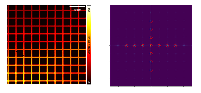
Jupyter notebook for the calibration of the XY-scanners of a microscope
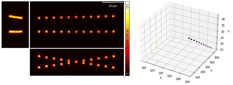
Jupyter notebook for the calibration of the Z-scanner of a microscope
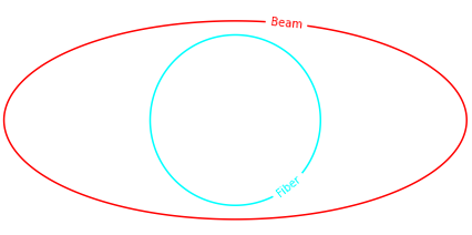
Jupyter notebook for the calculation of fiber coupling efficiency 
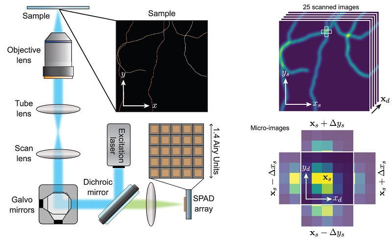
Video presentation on the basic concepts of ISM 
Published:
This is a description of your talk, which is a markdown files that can be all markdown-ified like any other post. Yay markdown!
Published:
This is a description of your conference proceedings talk, note the different field in type. You can put anything in this field.
Undergraduate course, University of Genoa, DIFI, 2019
Exercises on classical mechanics and electromagnetism.
Workshop, Istituto Italiano di Tecnologia, 2021
Instructor at the 6th, 7th, and 8th edition of the Advanced Microscopy practical workshop.
Ph.D. short course, University of Genoa, DIBRIS, 2025
Short course of the fundamentals of optics, imaging, and microscoppy.